


What is 3D planning in dentistry and what is it used for?
At Nemotec, we define 3D dentistry as the integration of diagnosis, planning and manufacturing around a virtual patient: a digital model that unifies bone, teeth and gums. With NemoStudio, we evaluate anatomy, simulate treatments and produce guides or splints with the surgical precision required in daily clinical practice.
What do we use it for?
- 3D dental diagnosis: we visualise bone volumes, roots and critical structures in detail.
- 3D planning: we simulate every detail before treatment to ensure maximum accuracy, safety and aesthetic results.
- Computer-aided manufacturing (CAD/CAM): we convert the plan into ready-to-use clinical devices (surgical guides, splints, provisionals and restorations).
How a 3D dental diagnosis works in a modern digital workflow
The Nemotec workflow is short, repeatable and measurable:
1. Data acquisition
-
- CBCT for bone, roots and anatomical structures (volume).
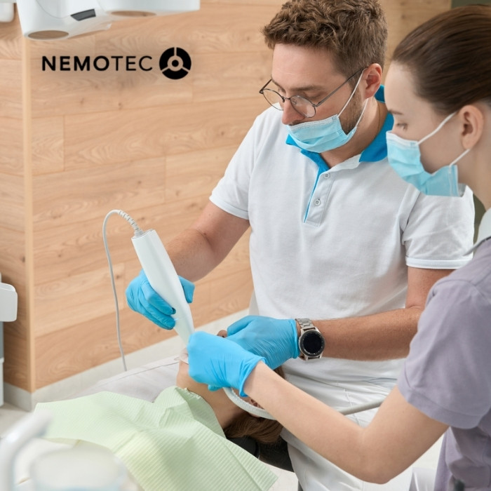
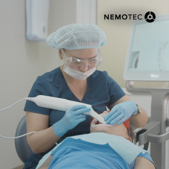
- Intraoral scanner for teeth and gums (surface).
Precision add-ons: integration of photographic records or facial scanning for comprehensive aesthetic analysis.
2. Fusion and control
-
- We merge CBCT (DICOM) with scans (STL/PLY/OBJ) into a single model.
- We validate the match in axial/sagittal/coronal slices to set the reference axis that will guide the entire treatment plan.
3. Collaborative 3D planning
-
- Orthodontics, implantology, surgery and aesthetics work on the same virtual patient.
- We version treatment proposals (v1, v2… v-final) with traceable comments.
4. Manufacturing
-
- We generate guides, splints and provisionals using prosthetic libraries and controlled parameters.
5. Execution and follow-up
-
- We implement the plan and record a verification scan, ensuring traceability and updating of the virtual patient.
Nemotec Experience: we use presets by case type (splints, pilot guides, anterior single implants, etc.) to reduce repetitive steps and ensure consistency.
Protocol we use
- Cleaning of CBCT artefacts when necessary.
- Export of the scan in STL (and PLY/OBJ if texture/colour is required).
- Registration using points and stable surfaces (palate, smooth surfaces, occlusal surfaces).
- Verification in three slices and record alignment.
Why 3D planning reduces clinical uncertainty and retreatments
- Everything in view: exact visualisation and measurement of the tooth-bone-gum relationship and precise matching with the patient’s photographic reality.
- Virtual rehearsal: surgical pathways, provisional occlusion and orthodontic sequences are validated before manufacturing.
- One language: clinic and laboratory share the virtual patient and comments in context.
- Quality control: checklists and versioning reduce rework and deviations.
Nemotec Experience: mandatory digital rehearsal (guide fit, occlusion and emergence profiles) before printing has consistently reduced retreatments caused by coordination mismatches.
Clinical applications of 3D dentistry in different specialties
3D planning in orthodontics
- Digital setup with traceable sequences and space/collision verification.
- TADs planned with adequate bone support.
- Decision between aligners vs. brackets based on anchorage, timing and complexity.
- Detailed workflow and tools in NemoFAB Ortho.
3D planning in implantology
- Prosthetically guided positioning based on the ideal emergence profile.
- Assessment of bone density and distances to critical anatomical structures.
- Use of real prosthetic libraries to avoid incompatibilities.
- Provisionals/immediate loading considering occlusion and tissues.
- Workflow described in digital implantology software.
3D planning in guided surgery
- Controlled pathways with sleeves and drills within known tolerances.
- Stability/support validated in the model (bone-, tooth- or mucosa-supported).
- From plan to print without intermediate reinterpretations.
3D planning in dental aesthetics
- Functional mock-ups (aesthetics + phonetics + occlusion) and preparation guides to transfer the design to the chair.
- Clear communication of the before/after in 3D to align patient expectations.
- Expanded methodology in 3D smile design software.
Limitations and best practices in digital dental planning
Limitations we address
- Input quality: a CBCT with artefacts or an incomplete scan compromises the entire workflow.
- Poor registration: if DICOM and scan data do not fit, clinical accuracy is lost.
- Outdated libraries: inconsistent connections and components lead to errors in guides/prosthetics.
Learning curve: without a protocol, the team spends more time than necessary.
Best practices we recommend
- Data collection: CBCT with an appropriate FOV; complete and recent scan.
- Fusion: validate in three slices and lock the reference.
- Versioning: v1-plan, v2-review, v-final (without overwriting).
- Libraries: same version in the clinic and laboratory.
- Digital rehearsal: check guide fit, occlusion and prosthetic space before printing.
- Internal metrics: plan acceptance, chair time, rework and number of appointments (continuous improvement).
Our operational recommendation: standard nomenclature (Patient_Surname_Date_Stage) and presets by case type to accelerate adoption and reduce human error.