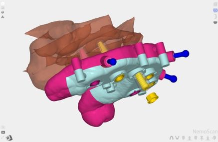
Dental implant planning doesn’t end when the clinician chooses the implant position on the CBCT. It ends when that position is transferred to the bone with enough precision that the prosthesis, the biomechanical load, and the relationship with adjacent structures are exactly what the plan anticipated.
Whenever an implant deviates from its planned position, something in the sequence becomes misaligned: the prosthetic emergence, the axial load axis, the bone margin, or the relationship with anatomical structures such as the dental nerve or the maxillary sinus. The clinical question is not whether there is a deviation, but which factors exacerbate it and which mitigate it.
In this article, we’ll explain what the evidence says about the factors that determine the precision of guided surgery, how we approach them when planning with NemoScan, and when it makes sense to outsource the case.
Why Implant Position Determines Everything
An implant is not a stand-alone piece. It is the starting point of a whole chain that includes the pillar, the crown, the relationship with the adjacent teeth, and the occlusal loads that the entire assembly will have to withstand for years to come.
The ideal position of the implant is not determined by the available bone, but by the final restorative outcome. The principle is reverse planning: first define where the prosthesis needs to be, then work backwards to the bone to establish where the implant has to go. When planning starts from the bone upward rather than from the final restoration, the prosthetic outcome is compromised before the patient even opens their mouth.
A 2 mm deviation at the entry point can turn an ideal prosthetic emergence into an unacceptable one. A 5° angular error shifts the loading axis off the implant. In the anterior sector, these errors affect the aesthetics; in the posterior sector, they affect the force distribution.
Digital planning doesn’t eliminate deviation: it quantifies it, anticipates it, and reduces it.
What CBCT Reveals Before Surgery That Radiography Can’t Show
Panoramic and periapical radiographs offer a two-dimensional projected image. For most decisions in implantology, they’re insufficient.
CBCT adds three layers of information that change clinical decisions:
- Actual bone volume in all three planes. Not just the height visible in the two-dimensional projection: the exact buccal-palatal or buccal-lingual width at the insertion point. Bone that looks adequate on a panoramic image can measure 4 mm wide on CBCT.
- Bone density and cortical quality. Density affects primary stability and the drilling protocol. A D3 or D4 bone area requires a different preparation sequence and may limit the viability of immediate loading.
- Precise relationship with critical structures. The distance to the inferior alveolar nerve, the maxillary sinus floor, or the roots of adjacent teeth is measured in real millimeters, not visual estimates. The recommended safety margin from critical structures is at least 2 mm.
Without CBCT, these parameters are estimated. With CBCT and planning software, they’re data.
The Factors That Determine the Precision of a Surgical Guide: What the Evidence Says
Guided surgery reduces intraoperative variability. But not all guides produce the same level of precision. Recent literature identifies four factors that determine how closely the executed result matches the digital plan.
The Type of Guide Support
Support is the factor with the greatest impact on final precision. Three recent systematic reviews agree on the same ranking:
Tooth-supported guide: the most precise. It rests on the remaining teeth, which offer a stable and reproducible reference. In the review by Marquez Bautista et al. (2024, Applied Sciences, Universidad Complutense de Madrid), covering 23 studies and 2,005 implants, the best results correspond to tooth-supported guides: coronal deviation of 0.39 mm, apical deviation of 0.28 mm, and angular deviation of 2.05°.
Bone-supported guide: precise, though somewhat more variable than the tooth-supported guide. It requires flap access to fix it to the bone.
Mucosa-supported guide: the most variable. It rests on soft tissue, which is compressible and offers less stability. In the reviewed studies, coronal deviation reaches 2.05 mm and angular error reaches 7.17° in the worst cases. Fixation with anchor pins improves its performance, especially in fully edentulous jaws.
The Type of Guidance: Fully Guided vs. Pilot-Only
The meta-analysis by Khaohoen et al. (2024, BMC Oral Health), which integrates 67 studies and 5,673 implants, quantifies the difference between the two protocols:
- Fully guided guide: controls direction, angulation, and depth throughout the drilling sequence.
- Pilot-only guide: controls depth and initial angulation, but the rest of the drilling sequence is freehand.
The difference is statistically significant (p<0.001): the fully guided guide reduces apical deviation by 0.44 mm and angular error by 3.29° compared to the pilot protocol. In implants near critical structures or in cases with planned immediate loading, that difference has a direct clinical impact.
Guide Fixation and Sleeve Design
Two design parameters affect execution precision:
Fixation pins: in mucosa-supported guides and in cases of total edentulism, fixation with anchor pins reduces movement during drilling. The recommendation is fixation at a minimum of three points; four points in fully edentulous jaws.
Sleeve-to-bone distance: the greater the distance between the guide’s sleeve and the bone crest, the greater the amplification of angular error at the apical point. Studies document worse precision when that distance exceeds 5 mm (Shi et al., 2023, International Journal of Implant Dentistry).
The Manufacturing Method
Guides produced by milling (CNC) show greater dimensional precision than 3D-printed guides, although the difference narrows when the printer works with calibrated resins under controlled conditions. In the review by Shi et al. (2023), manufacturing precision for milled guides ranges from 0.02 to 0.25 mm, while manufacturing precision for 3D-printed guides ranges from 0.03 to 0.44 mm. Both are clinically acceptable; milling shows less variability between cases.
How We Plan Dental Implants With NemoScan
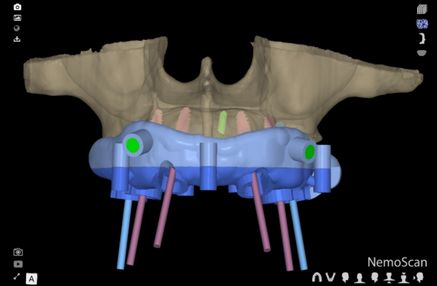
NemoScan integrates CBCT, models, 2D photography, and facial scanning into a single work environment. It covers the entire workflow from diagnosis to postoperative evaluation.
CBCT + Intraoral Scanner Fusion in a 3D Environment
The starting point is the integration of the two main records: the CBCT bone volume in DICOM format and the arch models in STL format. NemoScan overlays them to produce an accurate representation of the patient’s anatomy: bone, mucosa, teeth, and interarch relationships in the same space.
Case preparation is AI-assisted, which reduces segmentation time and increases the reproducibility of reference points.
Anatomical, Prosthetic, and Gingival Planning in the Same Plan
Implant planning in NemoScan integrates three dimensions simultaneously:
- Anatomical: available bone volume, density, proximity to critical structures.
- Prosthetic: implant position relative to the planned restoration. NemoScan allows visualization of the virtual wax-up and planning of the prosthetic emergence before designing the guide.
- Gingival: emergence profile, crest condition, and relationship with soft tissue.
For immediate implant cases, NemoScan allows virtual extractions to be performed for guide design.
Surgical Guide Design and Manufacturing
From the digital plan we generate directly:
- Pilot or fully guided guides: allow control of direction and depth and, in fully guided systems, control of the complete drilling sequence and implant placement.
- All-in-one guides: integrate tooth extraction, bone reduction when needed, implant placement, and adaptation of the immediate provisional restoration into a single system, reducing surgical time and improving the transfer of digital planning.
- GBR membrane designs: facilitate the positioning of membranes and/or biomaterials in cases with bone deficits that require simultaneous regeneration.
- Sinus lift guides: designed to guide the lateral window and surgical preparation in sinus lift procedures.
- Dental autotransplantation guides: allow the virtual planning of the recipient socket to be reproduced and facilitate positioning of the donor tooth, reducing extraoral time and helping preserve the periodontal ligament.
- Autologous bone graft harvesting guides: help delimit the volume and location of the graft at the donor site, increasing osteotomy precision and reducing the risk of injuring adjacent anatomical structures.
STL files are exported for in-house printing or sent to a lab.
Postoperative Verification: The Step That Closes the Loop
Once surgery has been performed, NemoScan allows the postoperative CBCT to be overlaid with the preoperative plan to quantify the actual deviation between what was planned and what was achieved. This step closes the case’s quality loop and provides objective information to refine subsequent protocols.
When to Outsource Implant Planning
Planning complex cases (fully edentulous, immediate loading, bone reduction, simultaneous GBR) has a real software learning curve. And clinic schedules don’t always allow the time needed.
From our guided surgery planning center, we offer the complete service:
- Complete implant planning
- Surgical guide design (pilot or fully guided)
- All-in-one guides
- Manufacturing or export of the guide’s STL files
- Other types of guides, such as guides for lateral sinus lift, autotransplantation, graft harvesting, and pathological lesion localization.
Frequently Asked Questions About Dental Implant Planning
What Records Do I Need to Plan Implants With NemoScan?
The minimum records are CBCT in DICOM format and arch models in STL format. Photography is optional but recommended for anterior cases where the emergence profile and lip relationship are part of the plan. Facial scanning can be added when the case’s esthetic component justifies it.
Can I Manufacture the Surgical Guide in My Own Clinic?
Yes. NemoScan generates the guide’s STL file directly from the plan. That file is compatible with any dental 3D printer and with biocompatible resins approved for intraoral use. If you’d rather not manufacture it in-clinic, our planning center includes manufacturing and shipping, or delivery of the print-ready STL file.
What’s the Difference Between a Pilot Guide and a Fully Guided Guide?
The pilot guide controls depth and angulation with a single drill; the rest of the drilling sequence is freehand. The fully guided guide controls direction, angulation, and depth throughout the entire drilling sequence. According to the meta-analysis by Khaohoen et al. (2024), the fully guided guide reduces apical deviation by 0.44 mm and angular error by 3.29° compared to the pilot protocol. In cases with a reduced safety margin or planned immediate loading, the fully guided protocol is recommended.
When Is Flapless Surgery Indicated?
Flapless surgery reduces trauma, shortens recovery, and cuts chair time. It’s appropriate when there’s enough keratinized gingiva, the available bone volume is sufficient without needing direct visual access, and the guide has stable fixation. When the case requires bone reduction, GBR, or visual assessment of bone volume, flap access is still necessary. Prior digital planning is what allows you to decide with confidence which approach applies to each case.
What Is an All-In-One Guide and When Should It Be Used?
All-in-one guides integrate tooth extraction, bone reduction when needed, implant placement, and adaptation of the immediate provisional restoration into a single system, reducing surgical time and improving the transfer of digital planning. They are especially useful for immediate post-extraction implants and full-arch rehabilitations where surgical efficiency is critical.
Do you have an implant case you want to plan with precision? Our team provides complete planning with delivery in just a few days. Contact our team.