


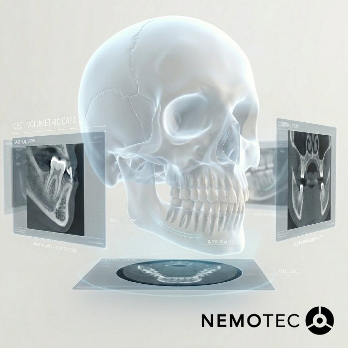
What is a dental CBCT and what information does it provide?
A CBCT (Cone beam computed Tomography) is a type of tomography that generates a 3D volume of the craniofacial region using multiple projections taken as the scanner rotates around the patient. In clinical practice, this volume is converted into multi-planar slices and reconstructions that are useful for diagnosis and treatment planning.
Nemotec´s experience: for us, the real breakthrough of CBCT is not just seeing in 3D, but being able to integrate that DICOM data with the rest of the records (STL/PKY from the intraoral scanner, JPG photos, even facial OBJ flies) and make decisions with the entire case within a single interface.
What does CBCT or cone beam mean in dentistry?
CBCT stands for Cone Beam Computed Tomography and is usually translated into Spanish as tomografía computarizada de haz cónico. It is a radiological technique that allows 3D images of the mouth and craniofacial structures to be obtained and is particularly useful for calcified tissues (teeth and bone).
What structures can be viewed in 3D: teeth, bone and nerves
It provides invaluable three-dimensional visualization for:
- Teeth (root morphology, relationship with impacted teeth, etc.)
- Alveolar and basal bone (volume, cortical bone, defects).
- Nerve canals and anatomical structures relevant to surgical safety.
What is a dental CBCT used for clinical practice?
Implantology and guided surgery
It us a cornerstone for:
- Assessing actual bone availability.
- Locating critical anatomical structures.
- Planning implants with prosthodontically guided approach.
- Feeding into guided surgery workflows (where applicable).
The Nemotec Experience: in implantology, what reduces complications most is not having a CBCT, but working with a consistent workflow + communication between clinic and laboratory. This unified workflow is precisely at the heart of NemoStudio.
If you want to see how a DICOM image is converted into an implant plan (position, angulation, depth and guide design), this is developed in the implant planning software.
Orthodontics, impacted teeth and bone analysis
In advanced orthodontics, CBCT helps to:
- Locate included/impacted teeth and their relationship with neighboring roots
- Assess bone limits (crucial if you want to plan safe movements)
- Improve diagnosis as we have all three spatial planes available.
Endodontics, TMJ and oral and maxillofacial surgery
In complex endodontics, oral and maxillofacial surgery, CBCT can assist with:
- Assessment of complex root anatomies.
- Suspected lesions that are not clearly visible in 2D.
- Study of sinuses, fractures, inclusions, and TMJ analysis in specific contexts.
How the scan is performed and what the patient should expect
How long it takes, how the patient should position themselves and whether preparation is required
On most machines, the scan is quick and is performed with the patient in a stable position (sitting or standing, depending on the device), with simple instructions to remain still. It does not usually require complex preparation beyond removing items that may cause artefacts (in accordance with your center’s protocol)
What happens after the scan and how are DICOM images interpreted?
The results are saved in DICOM format, which is the standard for visualization and planning using specialized software.
Nemotec´s experience: in our workflow, DICOM is not just a file; it is the starting point for 3D planning and, above all, for collaboration. When you can open, review and share the case with the team (clinic-laboratory-specialists), you reduce errors caused by isolated interpretation.
And when the aim is to scale up planning without adding more in-house hours, it makes sense to rely on a planning center to standardize criteria, review online and speed up deliverables whilst maintaining clinical control.
Differences between dental CBCT, CT and conventional radiography
When does it provide greater precision than a 2D scan?
2D imaging (periapical, bitewing, panoramic) remains useful, but CBCT offers clear advantages when you need to:
- Understand spatial relationships (vestibular-lingual) that 2D cannot resolve.
- Plan close to anatomical structures.
- Assess complex anatomies/impacted teeth.
Differences in field of view, detail and clinical utility
Here, the Field of View (FOV) is key:
- Larger FOV: greater anatomical coverage, but usually a trade-off in resolution and dose (depending on the protocol).
- Smaller FOV: more focused and efficient when the diagnostic objective is local.
Advantages of dental CBCT in diagnosis and treatment planning
Greater anatomical accuracy and improved decision-making
CBCT enhances three-dimensional anatomical assessment and, consequently, the quality of clinical decision-making in cases where 2D imaging is insufficient.
Lower radiation than a CT scan and greater clinical efficiency
Generally speaking, a dental CBCT typically involves a lower dose than a conventional medical CT scan, but the actual dose depends on the FOV, exposure parameters and equipment; therefore, the indication must be justified and optimized.
Integration with intraoral scanners and 3D planning software
The real value multiplier comes when you combine:
- DICOM (CBCT) + STL/PLY, JPG, OBJ)
- And you integrate it into a 3D planning environment.
At Nemotec this interoperability (DICOM, STL/PLY, JPG, OBJ) and Al-driven automation (e.g., segmentation) are designed to reduce pre-processing and free up time for clinical work.
Nemotec experience: when the team works with an open workflow (without “vendor lock-in”), you can update CBCT or IOS without disrupting your planning system. That continuity is what allows you to standardize protocols and scale up.
Limitations of CBCT and when it should be indicated on a case-by-case- basis.
Cases where it is not always the first test of choice
CBCT is not the ‘default’ option. It does not always add value over 2D imaging when:
- The diagnostic objective is adequately met with conventional X-rays.
- The case is straightforward and does not alter treatment decisions.
- The necessary FOV is not justified for the clinical question.
The importance of clinical judgement and the diagnostic objective
The indication must be based on a specific question: what clinical decision am I going to make with this information? Clinical guidelines (particularly in orthodontics) emphasize the need to justify the exposure and tailor the scan to the objective.
Frequently asked questions about dental CBCT
Is it the same as a dental CT scan?
In clinical conversation it is often referred to as a ´dental CT scan’, but CBCT and CT (medical CT) are not exactly the same: they differ in acquisition technology, standard protocols, and typical use in dentistry.
Does it emit a lot of radiation?
It depends on the equipment and protocol. In general, dental CBCT usually involves a lower dose than a medical CT scan, and the dose varies with the FOV and parameters, so it must be indicated and optimized on a case-by-case basis.
Does it hurt or have side effects for patients?
It is a non-invasive test: it does not hurt and leaves no residual `sensation’. What matters is the correct indication and the quality of the scan (to avoid repeats)
Is it useful for planning implants and guided surgery?
Yes: this is one of its most common uses, as it allows for planning with 3D visualization and exporting the scan in DICOM format for planning and guided surgery software (where appropriate).