TMJ disorders are not the exclusive territory of the orofacial pain specialist. Orthodontists planning a decompensation, prosthodontists designing a full rehabilitation, and surgeons scheduling an osteotomy all encounter active joint pathology more often than referral statistics suggest.
The problem is not a lack of knowledge about what temporomandibular disorders are. The problem lies in the gap between diagnosis and clinical decision-making: when the TMJ should shape the plan, when it should stop it altogether, and when it is safe to proceed under monitoring.
This article offers a clinical framework for approaching complex functional cases, from the minimum examination required before planning to the role of CBCT and the virtual articulator in integrating TMJ status into the digital diagnostic workflow.
Current Classification of Temporomandibular Disorders: The DC/TMD Framework
The Diagnostic Criteria for Temporomandibular Disorders (DC/TMD), developed by the International RDC/TMD Consortium, organize temporomandibular disorders (TMDs) into two complementary axes:
Axis I — Physical diagnoses, split into two categories:
- Arthrogenous: the disorder directly affects the joint. The most common entities are disc displacement with reduction (with or without clicking), disc displacement without reduction (with or without limited opening), arthralgia, osteoarthritis, and osteoarthrosis.
- Myogenous: no alterations are found in the joint complex, but muscular findings are present. Subtypes include local myalgia, myofascial pain (referred within the boundaries of the muscle examined), and myofascial pain with referral (extending beyond those boundaries).
Axis II — Psychosocial factors: degree of functional disability, and levels of anxiety, depression, and somatization, assessed through validated questionnaires (PHQ-4, SCL-90). The DC/TMD paradigm is biopsychosocial: it acknowledges that a single cause is unlikely to be identified in any given patient, and that Axis II factors modulate both pain threshold and treatment response.
In clinical practice, myalgia is the most frequent diagnosis, present in roughly 80% of TMD patients. Disc displacement with reduction is the most common joint finding; arthralgia and osteoarthritis are less prevalent but carry a greater impact on treatment planning.
Prevalence and Underdiagnosis in Specialist Practice
Prevalence data vary by population and diagnostic criteria, but the figures are consistently high. In the general adult population, estimates typically range from 20% to 40%. Studies in primary care push that figure higher: an analysis conducted in Chile using RDC/TMD criteria in 270 adults found that 49.6% met criteria for at least one TMD diagnosis, with 19.6% presenting severe functional limitation.
The gender distribution is consistent across the literature: women receive a TMD diagnosis in 61.3% of cases, compared with 30.4% of men. For treatment planning, this means that more than half of the patients undergoing orthodontic or prosthetic treatment carry a statistically significant likelihood of some degree of temporomandibular dysfunction.
Underdiagnosis in specialist practice has two main causes. The first is the absence of systematic screening before starting treatment: if the questions aren’t asked and the exam isn’t performed, mild joint or muscular findings never make it into the record. The second is subclinical presentation: many patients report no spontaneous joint pain, yet show signs of dysfunction that only surface on examination or that emerge once occlusal changes from treatment activate a joint that was already compromised.
The Clinical Examination That Should Precede Any Complex Treatment Plan
A TMJ examination requires no special equipment — only time and a protocol. The minimum components of the DC/TMD exam that every specialist planning complex cases should incorporate are:
- Maximum mandibular opening (assisted and unassisted): normal opening is 40 mm or more. Opening below 35 mm accompanied by pain suggests functional limitation.
- Joint sounds: clicking on opening or closing (suggestive of disc displacement with reduction), or crepitus (suggestive of degenerative bony changes).
- Opening and closing trajectory: lateral deviation on opening can indicate unilateral disc limitation.
- TMJ palpation: pain on lateral or posterior palpation under joint loading.
- Bilateral muscle palpation: temporalis (three sites), masseter (three sites), and the medial pterygoid region. Note whether pain is local or referred.
- Axis II screening: at minimum, one screening question on how pain affects daily activities, with referral to a validated questionnaire when there is suspicion of a significant psychosocial component.
The threshold for requesting advanced imaging is clinical: joint crepitus, limited opening that fails to improve after 4–6 weeks of conservative treatment, suspected inflammatory arthritis, progressive condylar asymmetry, or orthognathic surgery planning in a patient with a symptomatic TMJ.
CBCT in TMJ Diagnosis: What It Can and Cannot Tell You
What CBCT reliably detects
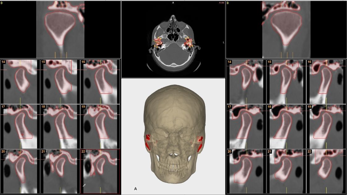
CBCT provides three-dimensional images with sub-millimeter resolution and no superimposition of structures. For the TMJ, it is the gold standard for assessing hard tissue:
- Condylar position within the glenoid fossa
- Degenerative bony changes: flattening of the condylar head, osteophyte formation, subchondral sclerosis, erosions
- Condylar asymmetry and ramus height discrepancies
- Radiographic signs of sustained parafunction
- Subcondylar fractures not visible on panoramic radiographs
What CBCT cannot tell you
The articular disc, ligaments, and synovial membrane are soft tissues that CBCT cannot visualize. To assess disc position and morphology, intra-articular inflammatory changes, or synovial effusion, MRI remains the gold standard. CBCT and MRI are complementary tools, not interchangeable ones.
CBCT as a triage tool
The role of CBCT in TMJ diagnosis is shifting from diagnostic confirmation toward triage identifying which patients need confirmatory MRI and which can be managed with bone imaging and clinical follow-up alone. A 2026 study published in Progress in Orthodontics (PubMed: 41661510) presents a deep-learning-assisted diagnostic model trained on CBCT to detect disc displacement in orthodontic settings. Its conclusion: this kind of tool can act as a filter that reduces reliance on manual expert interpretation and helps direct MRI referrals more efficiently.
Field-of-view protocol for TMJ CBCT
A large field-of-view CBCT exposes the patient to a higher radiation dose without improving resolution in the condylar region compared with a reduced field of view. For a focused joint evaluation, the recommended protocol is a bilateral, reduced field of view acquiring both joints in the same study, with slice thickness of 0.2–0.3 mm.
When the TMJ Blocks or Conditions the Treatment Plan
In orthodontics
An unstable TMJ is the most underestimated risk factor in the long-term prognosis of orthodontic treatment. When condylar position shifts during treatment whether from progression of active degeneration, resolution of a muscular dysfunction, or a change in the maxillomandibular relationship the occlusion built on the condyle’s initial position becomes misaligned.
The real risk: an orthodontic treatment executed flawlessly on a TMJ undergoing active degeneration can still end in an unstable occlusal relationship that is hard to trace back to any error in the plan. The distinction between stable dysfunction (reproducible condylar position, minimal symptoms) and active dysfunction (progressive changes, spontaneous pain, new-onset crepitus) is the clinical criterion that determines whether to stabilize first or proceed under monitoring.
In implant dentistry
Bruxism is the functional factor with the strongest documented impact on dental implant survival. It is not an absolute contraindication, but it is a plan modifier that calls for a specific protocol.
A 2026 systematic review following PRISMA methodology (Tarco-Rojas et al., YACHASUN) documents the most frequent mechanical complications in bruxist patients with implants: implant fracture, loosening or fracture of the prosthetic screw, abutment fracture, ceramic chipping, and prosthesis mobility. The recommended protocol includes preoperative bruxism screening (using the BruxScreen tool), prosthetic design with reduced cantilevers, selection of reinforced materials, a postoperative occlusal splint, and periodic occlusal follow-up.
In orthognathic surgery
Active condylar degeneration is a relative contraindication for orthognathic surgery. If the condylar head is losing bone volume during planning, the mandibular position being planned today may no longer be the correct one by the day of surgery, or six months later. Surgeries performed on actively degenerating joints show significantly higher relapse rates than those performed on stable joints.
The standard protocol is to stabilize the TMJ for at least 6–12 months before surgery, confirm with serial CBCT that condylar position is stable, and only then plan the definitive surgical movements.
Virtual Articulator and 3D Occlusal Analysis in Cases with TMJ Pathology
A conventional mechanical articulator mounts the models in a position recorded using a facebow and centric relation. That mounting assumes condylar position is stable and reproducible. In a patient with active TMD, that assumption is questionable: if condylar position shifts with muscle pain, intra-articular edema, or the opening cycle, the mounting captures a single moment, not a stable reality.
The virtual articulator addresses this from two angles. The first is reproducibility: the digital model can be remounted at different condylar positions with no material or time cost. The second is CBCT integration: the patient’s actual condylar position, documented through imaging, transfers directly into the digital environment without depending on the technician’s precision during physical mounting.
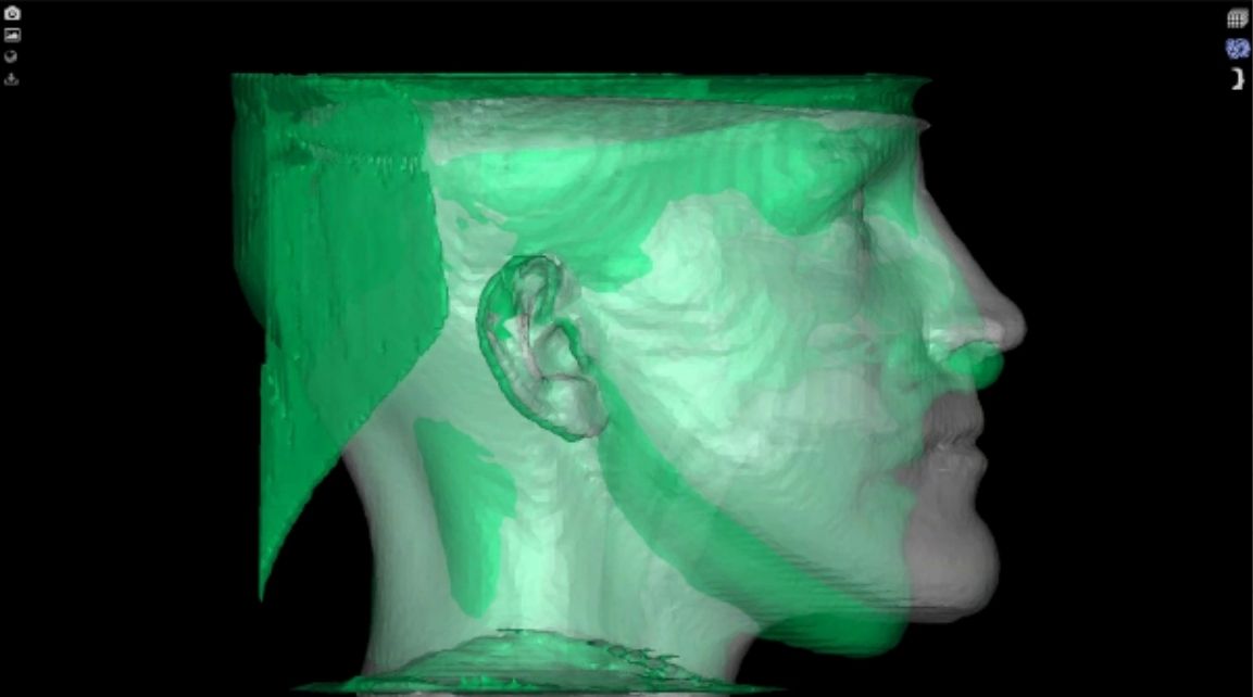
Integrating CBCT, intraoral scans, and facial scans within a virtual articulator makes it possible to analyze mandibular kinematics detecting premature contacts in excursive movements, evaluating anterior guidance, and checking whether the planned vertical dimension creates interferences, all before a single provisional is fabricated.
NemoFAB Ortho integrates TMJ analysis directly into the digital planning workflow. Joint status becomes part of the diagnosis alongside 3D cephalometry, airway analysis, and occlusal planning, all within a single environment. In cases combining TMJ dysfunction with complex orthodontic or prosthetic planning, this level of integration reduces the number of decisions the clinician has to make with incomplete information.
Treatment Sequencing in Cases with Active TMD
The literature is unanimous on one point: no irreversible occlusal treatment should begin on an unstable TMJ. Starting crowns, implants, or surgery on a joint in active dysfunction is the root cause of some of the hardest-to-resolve failures in oral rehabilitation.
The recommended sequence follows four phases:
- Reversible stabilization. A stabilization occlusal splint (Michigan-type or similar), with progressive adjustment. The goal is not to eliminate the TMD but to bring the joint to a reproducible position with controlled symptoms. Duration ranges from 4 weeks to 6 months depending on severity.
- Updated diagnostic imaging. Bilateral TMJ CBCT taken in the stabilized position. If clinical examination suggests active disc pathology, MRI to assess soft tissue. The prosthetic or orthodontic plan should be built on this imaging, not the initial one.
- Integrated 3D planning. With the stabilized condylar position documented, the virtual articulator allows occlusal movements or skeletal changes to be planned with reference to the patient’s actual TMJ.
- Definitive treatment. Only once the previous phases are complete and the joint is stable. Follow-up afterward should include periodic evaluation of joint status, not just the occlusal outcome.
Frequently Asked Questions About TMJ Disorders
What’s the difference between joint clicking and TMJ arthralgia?
Joint clicking is a sound produced by disc displacement and reduction during mandibular opening or closing. On its own, it does not indicate progressive joint damage many patients have clicking with no symptoms for years. Arthralgia is joint pain on palpation or movement, and does not require the presence of any sounds. The distinction matters for planning because painless clicking rarely conditions the start of treatment, whereas active arthralgia does.
Can a patient with active TMD start orthodontic treatment?
It depends on the type and stage. Mild, stable, asymptomatic muscular dysfunction does not contraindicate orthodontics. Active condylar degeneration, persistent arthralgia, or disc displacement without reduction with limited opening do condition the start of treatment the correct sequence is to stabilize first and plan afterward. The most common mistake isn’t treating patients with TMD; it’s starting treatment without knowing the dysfunction is there, or without adapting the plan to it.
Does bruxism contraindicate dental implants?
Not absolutely. Bruxism is a documented biomechanical risk factor that increases the incidence of mechanical complications (implant fracture, screw loosening, abutment fracture, ceramic chipping), but the available studies do not show significantly higher osseointegration failure rates when the case is properly protocolized. Bruxism does modify the plan: reduced cantilevers, material selection, strict occlusal control, and a postoperative splint are part of the standard protocol in these cases.
What minimum records are needed before planning a case with suspected TMJ pathology?
The DC/TMD clinical examination is the starting point: mandibular opening, joint sounds, and bilateral joint and muscle palpation. If the exam suggests active pathology, a reduced-field-of-view bilateral TMJ CBCT is the next step for assessing bone tissue. If there is suspicion of active disc displacement, limited opening, or persistent arthralgia, MRI completes the soft-tissue diagnosis. The virtual articulator combining CBCT, STL models, and a centric relation record taken in the stabilized positioncloses out the functional diagnosis before any definitive treatment is planned.
What does the virtual articulator offer over the mechanical one in cases with TMJ dysfunction?
A mechanical articulator depends on the accuracy of the centric relation record and the technician’s mounting. In patients with active TMD, where condylar position shifts with pain or intra-articular edema, that record may not be reproducible. The virtual articulator allows the clinician to work with the condylar position documented via CBCT, remount it at no material cost, and analyze occlusal dynamics in real time. It also facilitates communication between clinician and lab, and supports longitudinal documentation of the case.
Have a complex case with TMJ pathology that needs to be integrated into your digital planning? NemoFAB Ortho brings TMJ analysis, the virtual articulator, and 3D cephalometry together in a single workflow. Check out the Nemotec planning center for outsourced cases.