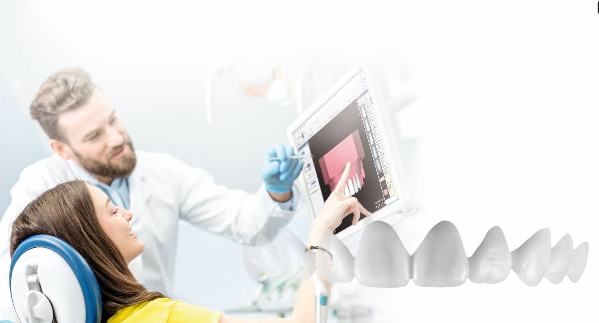
In the clinic, the comparison of “CBCT vs CT” is not about technology out of curiosity. It is about what information we need, at what level of detail, in which anatomical region and at what operational and radiological cost. If we choose correctly, we gain diagnostic predictability and 3D planning without adding unnecessary complexity—or exposure.
And here is the important part: CBCT stands out when the objective is dentomaxillofacial and the clinical decision depends on millimetres (bone, teeth, roots and their relationship with anatomical structures). CT (medical CT) usually comes into play when we need a broader anatomical scope, hospital protocols or an evaluation that goes beyond the dental setting.
What is CBCT and how does it differ from CT in dentistry?
CBCT (Cone Beam Computed Tomography) is a tomographic technique that uses a cone-shaped beam and reconstructs a 3D volume that is particularly useful for dentomaxillofacial structures. CT (in its usual medical use) employs fan-beam technology and protocols designed for a broader anatomical spectrum.
In dentistry, the practical difference is usually this: CBCT prioritises spatial resolution and control of the field of view (FOV) in the area of interest, while CT tends to prioritise coverage and contrast according to the medical indication.
CBCT vs CT: key differences every dental clinic should know
Beam type, image acquisition and reconstruction
- CBCT: cone beam and detector, volume acquisition and 3D reconstruction focused on the maxillofacial region.
- CT: fan-beam technology with slice-by-slice reconstruction, commonly used in medical radiology settings.
In daily practice, this means that CBCT generally provides a volume better suited to teeth and jaws, with a more direct workflow towards dental planning.
Radiation, field of view (FOV) and resolution
Here are two rules we always apply:
- The FOV should be adjusted to the clinical question.
The further you go beyond the necessary area, the more exposure and diagnostic noise you introduce. In dentistry, using a larger FOV “just in case” is often a mistake. - Lower radiation does not mean indiscriminate use.
The indication is what matters. The aim is to optimise the minimum necessary to answer the clinical question safely.
In technical terms, CBCT usually offers spatial resolution that is highly useful in dentomaxillofacial applications. CT generally performs better in situations where the indication requires medical protocols and greater coverage.
Patient positioning, acquisition time and operating cost
There is significant variation depending on the equipment and protocol, but operationally three patterns tend to emerge:
- CBCT is easily integrated into the dental workflow (scheduling, planning and internal referral).
- CT usually depends on a radiology or hospital pathway and more general protocols.
- The real total cost is not only the scan itself; it also includes potential repeat scans due to incorrect indication and time lost through lack of coordination.
What advantages does CBCT offer over CT in dental practice?
Greater precision for dentomaxillofacial structures
If you work in implantology, advanced orthodontics or complex endodontics, you know why: CBCT gives you a 3D assessment that fits dentoalveolar structures particularly well when the clinical question is local and millimetric.
Lower exposure in many scenarios and greater control of the area studied
In dental settings, comparative evidence tends to show that CBCT may involve a lower effective dose than medical CT, especially when the FOV is well selected and the protocol is optimised.
In addition, FOV control avoids paying—in exposure and diagnostic noise—for anatomy that is not needed.
More useful measurements for clinical planning and communication
This is where we take a strong position: a CBCT should not remain in a viewer. It should become operational data.
When we turn DICOM into part of 3D planning and integrate it with the rest of the records, the conversation changes: we move from looking to deciding. To apply this in implantology and guided surgery, the natural fit is NemoScan (CBCT-based planning and guided workflow).
Uses of CBCT in dentistry according to specialty
Orthodontics and aligners
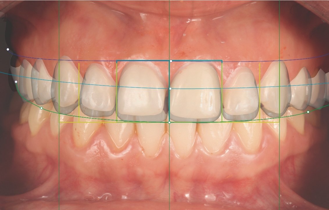
In orthodontics, CBCT adds value when we need to understand biological limits (roots/bone) and make safe decisions in complex movements. When we combine it with digital models, the case stops being just an image and becomes a coherent 3D plan, as with NemoCast 3D.
Implantology and guided surgery
In implantology, CBCT is often the logical starting point when planning is prosthetically guided and guide fabrication is required. The real step forward occurs when the workflow is seamless between clinic, planning and laboratory.
If the clinical team wants to scale complex cases without slowing down production, the most efficient option is to rely on a planning service with online validation. That is why we have our Planning Centre.
Prosthetics, veneers and rehabilitation
In rehabilitation, the value appears when we unify records: CBCT (DICOM) + intraoral scan (STL/PLY) + photographs (JPG). This integration reduces uncertainty and avoids blind decisions caused by fragmented data.
Endodontics, TMJ and pathology
In endodontics, TMJ and pathology, the indication must be especially precise: a clear clinical question, an adjusted FOV and responsible interpretation. CBCT contributes, but it does not imply unlimited use.
When is CBCT appropriate and when might CT make sense?
Cases in which CBCT is usually the most appropriate option in a dental clinic
- Local dentoalveolar questions where you need 3D imaging with a controlled FOV.
- Treatment planning where 3D imaging will be integrated with digital models and planning.
- Cases where optimisation and justification allow the study to be limited to the region of interest.
Cases in which CT may be more appropriate due to anatomical scope or hospital referral
- Need for broad anatomical coverage or hospital protocols.
- Cases requiring coordination with medical radiology and assessment beyond the dental setting.
- Situations where the clinical indication is not purely dentomaxillofacial.
How to integrate CBCT into a dental digital workflow
Combining CBCT, intraoral scanners, photography and STL/DICOM files
Our way of working is straightforward: if the data does not flow, the 3D remains on the screen.
That is why we work with an ecosystem that supports and integrates records in standard formats: DICOM (CBCT) + STL/PLY (intraoral scanning) + JPG (photography) + OBJ (facial scanning), eliminating silos between specialties and facilitating clinical-laboratory collaboration.
3D planning, guided surgery, aligners and prosthetics
When these records coexist, planning becomes actionable: you reduce improvisation, increase predictability and reduce chair time by moving decisions into the virtual environment.
Compatibility with open software and multidisciplinary ecosystems
Here we are very direct: the biggest risk of a digital investment is vendor lock-in. That is why we advocate open ecosystems, where you can evolve your technology stack without rebuilding your operational core.
Common mistakes when comparing CBCT and CT in a dental clinic
Confusing lower radiation with indiscriminate use
The fact that a protocol may involve a lower dose does not mean it should always be used. International guidance insists on justification and optimisation (clinical question + parameters + FOV).
Choosing a larger FOV than necessary
This is the silent mistake: you expand the FOV “just in case” and end up exposing and analysing anatomy you did not need. Adjusting the FOV to the indication is an explicit recommendation.
Assessing the equipment without considering workflow and training
The return does not come from the equipment alone. It comes from:
- protocol + indication
- data integration
- and a trained team
If you want to validate compatibility and implementation of the workflow, including CBCT/DICOM, we make it easy with our technical requirements, so that the workflow does not lose efficiency because of an operational detail.
Frequently asked questions about CBCT vs CT
Does CBCT always replace CT?
No. In dentomaxillofacial cases it is often very efficient, but there are scenarios where CT makes sense because of anatomical scope, protocol or hospital referral.
Which test is suitable for implants, orthodontics or endodontics?
It depends on the clinical question and the region. For implants and guided surgery, CBCT is common when the plan requires 3D integration with digital models. For orthodontics and endodontics, the indication should be refined case by case, with an adjusted FOV.
How do FOV and resolution affect the indication?
The FOV determines which anatomy is included—and exposed. The recommendation is to adapt it to the indication and avoid exceeding what is needed.
Resolution (voxel size) affects the useful level of detail, but also noise and acquisition parameters: “more resolution” is not always better if it does not answer the clinical question.
What should a clinic assess before incorporating it?
- Which clinical decisions need to be supported with 3D.
- How DICOM will be integrated with STL and photographs so that the data is useful.
- The protocol for indication, optimisation and FOV selection, with genuine justification.
- Team training and support for complex cases.