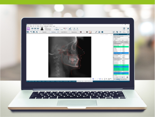
What is a cephalometric analysis in orthodontics?
A cephalometric analysis is the process of tracing, measuring and interpreting craniofacial landmarks (points, planes, angles and proportions) to describe skeletal and dentoalveolar relationships, estimate growth patterns and support treatment decisions.
In a digital practice, analysis ceases to be a collection of numbers and averages that we sometimes overlook, and instead becomes the link that connects our records: CBCTs, intraoral scans (STL/PLY) and photographs, to ensure consistency in diagnosis and facilitate interdisciplinary planning. That is why, within NemoStudio, we develop this module using tools such as NemoCeph.
What is its purpose in clinical diagnosis?
The clinical value lies in reducing uncertainty: identifying discrepancies, prioritising objectives and anticipating trade-offs.
Skeletal and dentoalveolar assessment
- Sagittal skeletal diagnosis: maxillomandibular discrepancy and its relative weight compared to dental compensation.
- Vertical assessment: facial pattern, rotations and their impact on mechanics, anchorage and stability.
- Dentoalveolar component: incisor inclinations, protrusion/retrusion and degree of compensation.
From an operational perspective, the latent risk lies not in the measurement itself, but in the lack of clinical application of these measures due to a lack of integration with other records.
Orthodontic and surgical planning
- Determine whether the case is a candidate for orthodontic camouflage or requires a surgical approach (depending on skeletal discrepancy, biological limits and aesthetic-functional objectives).
- Define cephalometric objectives (and, where appropriate, VTO) consistent with the overall plan.
- In orthognathic cases, align the analysis with 3D planning to ensure consistency between objectives, treatment delivery and the final result.
When a case requires advanced planning or operational scalability, we offer support through the planning centre, enabling the outsourcing of part of the technical planning whilst ensuring that clinical control remains with the practitioner.
How cephalometric analysis is performed
The robustness of the analysis depends on two variables: the quality and standardisation of the records and the consistency of the tracing.
Required radiographic and digital records
Baseline (2D):
- Standardised lateral teleradiograph (and PA if your protocol includes it).
- Clinical photograph for aesthetic context and posture assessment.
Extended (2D/3D):
- CBCT in DICOM (where indicated) + intraoral scan (STL/PLY) + photograph (JPG) and/or facial scan (where applicable). This approach allows us to work with a more comprehensive set of data and reduces errors arising from partial assessment. In NemoStudio, this is supported by compatibility with open formats and an interoperable environment.
Identification of cephalometric landmarks
Recommendations for intra- and inter-observer consistency:
- Standardise the set of landmarks and the analysis method (Steiner/Ricketts/McNamara/Arnett or another) according to case type.
- Define explicit rules for landmarks that are difficult to identify (overlaps, diffuse cortical margins).
- Implement internal audits and correction protocols when deviations are detected.
Interpretation of planes, angles and proportions
To avoid a numerical reading without clinical interpretation, we recommend the following sequence:
- Skeletal sagittal (maxillomandibular relationship).
- Vertical (facial pattern and rotations).
- Dentoalveolar (compensations, incisors).
- Soft tissues (if included in the protocol).
- Clinical conclusion: treatment objectives, limitations and risks.
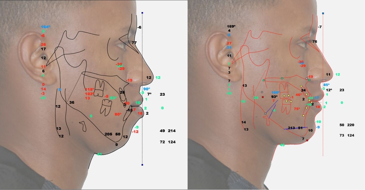
What parameters does a cephalometric analysis measure?
The value of each parameter lies in its ability to influence a clinical decision.
Maxillomandibular relationship
- Indicators such as SNA/SNB/ANB and/or Wits (depending on the protocol).
- Clinical interpretation: extent of discrepancy or compensation; implications for mechanics, extractions, distalisation, etc.
Facial pattern and growth
- Measurements associated with divergence/rotation (e.g., the mandibular plane in relation to cranial landmarks).
- Implications: vertical control, stability, management of open/closed bites and induced mandibular rotations.
Tooth inclination and compensations
- Inclination and position of the incisors in relation to the bony bases.
- Identifying compensations: when they are acceptable (camouflage) and when they compromise periodontal limits or stability, particularly if confirmed by 3D imaging where appropriate.
Digital cephalometry and AI in clinical practice
The aim is not more technology, but greater consistency, less unproductive time and better coordination.
Automatic landmark tracing
AI can improve the efficiency and reproducibility of the initial tracing, particularly in high-volume clinics. Our operational recommendation:
- Identify critical landmarks (“red flags”) that are always checked manually.
- Establish a validation checklist before finalising the diagnosis.
Within the NemoStudio ecosystem, the automation of tracing/segmentation is described as a mechanism for optimising clinical time, whilst maintaining the professional’s role in the final review.
Integration with CBCT, STL and 3D planning
The integration of DICOM + STL/PLY + photography enables:
- Correlation between cephalometric diagnosis and actual anatomy (roots, alveolar bone, asymmetries).
- Improved interdisciplinary planning (orthodontics, surgery, implantology, aesthetics) with a unified view of the case.
Benefits in terms of efficiency, accuracy and interdisciplinary communication
- Operational efficiency: fewer repetitive manual tasks and less friction in the exchange of information.
- Practical accuracy: reduced errors due to fragmentation; consistency between diagnosis and treatment planning.
- Clinical communication: traceability of decisions and team alignment, useful in referrals and committee meetings.
Limitations and interpretative criteria
Anatomical variability and quality of the record
Cephalometric analysis is influenced by:
- Individual anatomical variability.
- Quality of the record (posture, definition, superimposition).
- Changes due to growth and functional adaptation.
Practical guideline: in the event of inconsistencies, review the records and the tracing before “adjusting” the interpretation.
Why software cannot replace clinical judgement
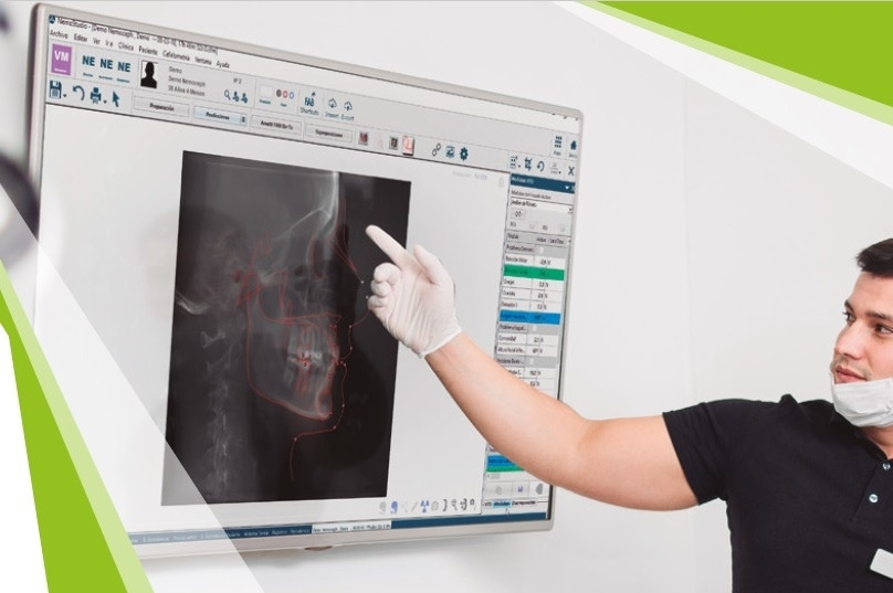
Software provides speed and standardisation; diagnosis requires clinical judgement: the integration of records, treatment objectives, risks and biological limits.
In our approach, technology serves as a decision-support infrastructure, not as a substitute for the professional, prioritising predictability and efficiency without shifting clinical responsibility.
Frequently asked questions about cephalometric analysis
What is the difference between 2D and 3D cephalometry?
2D (teleradiography) is standardisable and efficient; 3D (CBCT) provides volume and anatomical context where indicated. Optimal performance is achieved by integrating DICOM with STL/PLY models and photography within a unified workflow.
Which cephalometric points are most critical?
Those that most influence decisions: A/B, pogonion/menton, incisors and key planes of the protocol used. We recommend defining an internal critical set and auditing consistency.
What does AI contribute to cephalometric tracing?
It enhances efficiency and standardises the initial tracing. The clinical standard is to automate and verify, particularly for landmarks with high diagnostic sensitivity.
When should a cephalometric analysis be repeated?
According to protocol and clinical judgement: changes in growth phase (start/end), significant growth, or the need to reassess biomechanical or surgical hypotheses.
How does it integrate with aligner planning?
It integrates as a diagnostic layer (sagittal/vertical/incisor limits) and is enhanced in 3D workflows by correlating with STL/PLY and DICOM files where appropriate, particularly in complex movements where roots and bone play a decisive role.
Which errors affect cephalometric diagnosis the most?
In practice, the errors that most distort cephalometric diagnosis usually originate before any measurements are interpreted. The first is poor recording, whether due to an unstable posture, a poorly standardised projection or insufficient definition of structures, which introduces biases from the outset. From there, inconsistency in the identification of landmarks is common, especially at points with anatomical overlap or indistinct borders: small variations at these points generate significant changes in planes and angles, and therefore in the clinical interpretation. Another common error is falling into an exclusively numerical interpretation, without correlating the findings with the examination, photography and models, and, when appropriate, with 3D information. This can lead to conclusions that are correct “on paper” but incorrect for the patient. Finally, in digital workflows, a real risk is relying on automated processes without professional validation: automation can improve efficiency, but diagnostic closure always requires review and clinical judgement, especially regarding the points and measurements that influence therapeutic decisions.