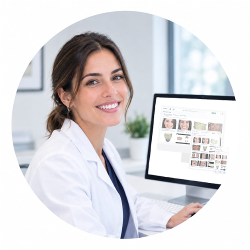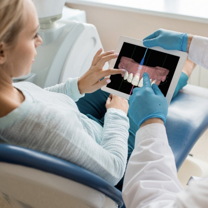
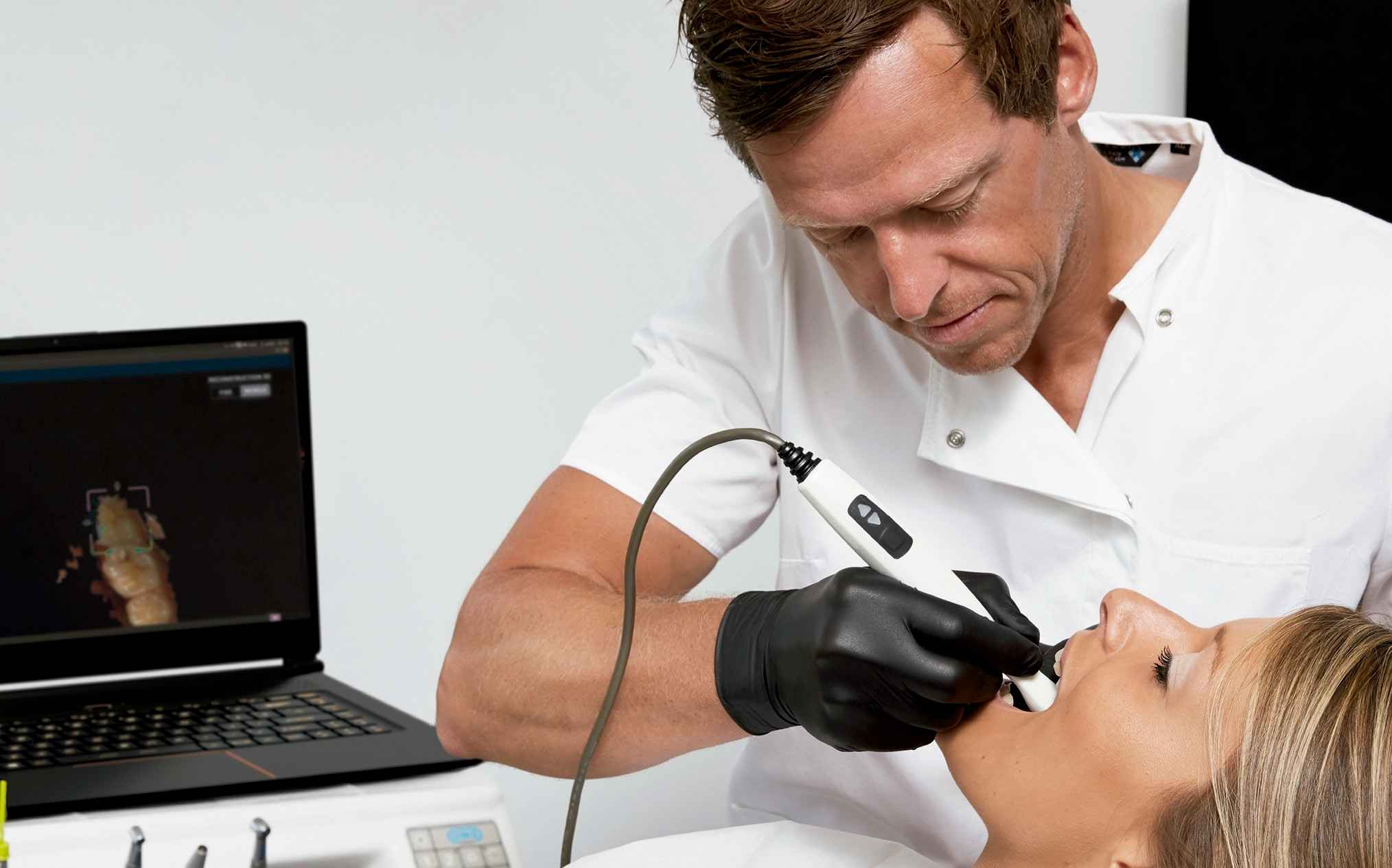
In the clinic, the intraoral scanner (Intraoral Scanner, IOS) is the device that allows us to capture dental anatomy in 3D directly in the mouth and turn it into a digital model ready for diagnosis, planning and manufacturing.
For us, the intraoral scanner is not just an extra: it is the entry point for data. When the data is captured correctly—clean, complete and verifiable—everything that follows (orthodontics, implantology, prosthetics and cosmetic dentistry) becomes more predictable and more efficient. To place it within the overall workflow, here we explain what NemoStudio is and why it works as the core of an interdisciplinary digital workflow.
Difference between an intraoral scanner, a conventional impression and a digital model
- Conventional impression: we capture the anatomy using materials and a tray, obtaining an impression and then a physical model. It is a valid process, but it introduces variables such as deformation, timing, transport and casting.
- Intraoral scanner: the record becomes an immediate 3D model that we can review on the spot, minimising uncertainty.
- Digital model: it is the final asset: a 3D file that is measurable, shareable and integrable within a CAD/CAM workflow.
In our experience, the real change is not simply going digital. The change is stopping working in disconnected parts: when we unify records and specialties in an interoperable environment, we reduce clinical-laboratory friction and avoid information silos.
How an intraoral scanner works step by step
Real-time 3D image capture
The IOS performs multiple captures and stitches them together to build the 3D geometry. Here, the decisive factor is not just the hardware: it is the clinical protocol.
In the clinic, we treat it as a diagnostic record: isolation, saliva control, selective drying and a stable scanning path. This discipline produces models that are both visually accurate and clinically reliable.
Digital file generation and immediate review
A major operational advantage is that we validate immediately: incomplete areas, stitching errors, occlusal inaccuracies and unclear margins can be seen and corrected before exporting. This reduces repeat scans and shortens the cycle with the laboratory.
Sending to the laboratory or CAD/CAM planning software
The value of the IOS increases significantly when the file enters a workflow that allows us to integrate it with the rest of the records. For this reason, we work with an open ecosystem capable of handling standard intraoral scan formats (STL/PLY) and combining them with CBCT (DICOM), clinical photography (JPG) or facial scanning (OBJ).
Which clinical cases is it used for?
Orthodontics and aligners
Intraoral scanning is the basis of digital diagnosis and treatment setup. When we also work with 2D/3D planning, we gain control and safety in complex movements. For orthodontic diagnosis and planning, we support the workflow with tools such as NemoCeph (cephalometric diagnosis) and, when the case requires it, digital orthodontic and production modules.
When the objective is to work with models, setups and digital products such as aligners and guides, NemoCast fits naturally as part of the ecosystem.
And when more advanced orthodontic planning is required—TMJ analysis, dynamic occlusion analysis and 3D cephalometry—NemoFab Ortho is the next logical step.
Implantology and guided surgery
In implantology, the IOS is essential for moving from implant placement to prosthetically guided planning. The objective is to align anatomy, emergence profiles, occlusion and guide fabrication within a coherent workflow.
Prosthetics, veneers and rehabilitation
In rehabilitation and cosmetic dentistry, intraoral scanning optimises communication with the laboratory: capture, design, validation and manufacturing. When properly protocolised, it supports more efficient communication and increases consistency of the final result.
Smile design and patient communication
3D visualisation helps to explain and align expectations. When a clinic approaches aesthetics systematically, NemoSmile enables design and communication through a digital workflow focused on predictability.
Advantages of the intraoral scanner for the clinic and the patient
Greater precision and fewer repeat scans
The practical benefit is clear: by verifying immediately, defective records that require repetition are reduced, decreasing time loss and clinical-laboratory friction.
Greater comfort and a better patient experience
Fewer materials, less setting time and a better experience for the patient. In the clinic, this translates into greater cooperation and speed.
Time savings in the clinic and laboratory
There are fewer intermediate steps. When we unify data in a common architecture, we also reduce duplicated planning and repetitive tasks.
Better treatment acceptance through 3D visualisation
Visualisation is not just “presentation”: it is uncertainty reduction. When the patient understands the plan, the process moves faster and adherence improves.
With Nemo Smile Generation, the patient can visualise their future smile through ultra-realistic images and videos powered by artificial intelligence. These tools use an intraoral scan and previous photographs, which are transformed into a powerful visual resource to improve understanding of the case and increase acceptance of treatment plans.
Limitations and when it does not fully replace conventional impressions
Full arches, soft tissues and other demanding cases
There are particularly demanding scenarios, including full arches, soft tissue control, bleeding, subgingival margins and hard-to-access surfaces. In these cases, the IOS may require a stricter protocol and, at times, does not replace a conventional impression completely depending on the clinical objective.
Common scanning errors and how to avoid them
These are the most common issues we see, and how we prevent them:
- Moisture and saliva → isolation and selective drying.
- Unstable scanning path → a fixed and repeatable scanning pattern.
- Bright areas/reflections → drying and lighting control.
- Lack of references → capture anatomical structures first, then smooth areas.
- Failure to validate occlusion and margins → a review checklist before exporting.
How it fits into a complete digital workflow
From intraoral scanning to CBCT, photography and 3D design
The IOS is one piece. Complete digital dentistry is achieved when you integrate STL/PLY (intraoral), DICOM (CBCT), clinical photography (JPG) and facial scanning (OBJ) in an environment where anatomy can be viewed as a whole and planned with interdisciplinary consistency.
Integration with open software and compatible hardware
In our architecture, the key is to avoid vendor lock-in: the clinic must be able to update hardware without rebuilding its operational core. That is why we advocate interoperability.
From diagnosis to manufacturing: guides, aligners and prosthetics
When the record is reliable and the treatment plan has been validated, the workflow leads to manufacturing: guides, splints, aligners and prosthetics. The objective is simple: clinical predictability + operational efficiency.
Frequently asked questions about the intraoral scanner
Does it hurt or feel uncomfortable for the patient?
It should not hurt. It can be uncomfortable for patients with limited opening, a gag reflex or high salivation, but when properly protocolised it is generally more tolerable than conventional impressions.
Is it suitable for orthodontics, implants and prosthetics?
Yes. The key is the case and the integration of records to plan safely, particularly when combining intraoral scans with CBCT and photography.
What files does it generate and how are they shared?
In open workflows, we typically use STL/PLY for intraoral scans and integrate them with DICOM (CBCT) and other formats (JPG/OBJ) depending on the case. Files are shared by export or through cloud environments and web viewers when the ecosystem allows it.
What should a clinic assess before incorporating it?
- Type of cases (orthodontics/implants/prosthetics/aesthetics).
- Laboratory and real compatibility.
- Internal protocol and team training.
- That the scanner fits into a complete digital workflow and does not remain an isolated tool.